Choose your shipping method in Checkout. Costs may vary based on destination.
Seller's Description:
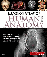
Very good in very good dust jacket. Junge Ärzte stehen vor der Aufgabe, den gezielten Einsatz der modernen bildgebenden Methoden zu trainieren und die damit erhobenen Befunde sicher zu interpretieren. Dies erfordert die genaue Kenntnis der Anatomie des Menschen, so wie sie sich in diesen Verfahren darstellt. Der Atlas von Professor Jamie Weir und Dr. Peter H. Abrahams stellt in 600 Abbildungen die normalen anatomischen Verhältnisse des Menschen in den bildgebenden Verfahren dar. In Zusammenarbeit mit sechs weiteren Experten auf dem Gebiet der Neuroradiologie und Radiologie wurden die Bilder mit Hilfe modernster Technik gewonnen und die anatomischen Strukturen identifiziert und beschrieben. Anatomische Details sind im Bild mit Zahlen markiert und so mühelos in den ausführlichen Bildlegenden identifizierbar. Auf Computer-und Magnetresonanztomogrammen können spezifische Strukturen in aufeinanderfol2enden Schichtebenen verfolgt werden. Besonders strukturreiche anatomische Regionen werden zum besseren Verständnis mit Hilfe verschiedener bildgebender Verfahren dargestellt. Dem Arzt ist der Atlas eine Entscheidungshilfe für die Auswahl des richtigen diagnostischen Verfahrens und die Abgrenzung pathologischer Veränderungen von normalen Verhältnissen. Dem Medizinstudenten hilft er, das im Anatomiekurs gewonnene Wissen zu vertiefen und auf die Diagnosefindung in der klinischen Praxis anzuwenden.